Photographing an Invisible Frog and what it Reveals About Blood ... - USC Viterbi School of Engineering

The translucent abdominal skin of the glass frog forms part of its unique disguise. Image/Geoff Gallice
How can you capture a detailed image of an invisible thing?
This is the challenge that a research team in USC's Alfred E. Mann Department of Biomedical Engineering had to overcome.
The results enabled a groundbreaking multi-institution investigation into the behavior of glass frogs — small, tree-dwelling, clear-skinned amphibians that can evade predators while sleeping. How? By making themselves invisible.
The research, recently published in Science, was led by scientists at Duke University, Dr. Junjie Yao, Dr. Sonke Johnsen, and Dr. Lauren O'Connell from Stanford University, with the USC collaborative team led by Professor of Biomedical Engineering and Ophthalmology Qifa Zhou.
The Zhou Lab is renowned for developing advanced ultrasound transducer and imaging techniques. Zhou pioneered the first-ever high-sensitivity single crystal transducers for ultrasound and photoacoustic imaging. The system has critical applications for cancer imaging, as well as imaging within the brain, blood vessels, and eyes. Zhou was approached by his Duke University collaborator Yao who was looking for advanced photoacoustic technology to study glass frogs, which can provide a unique insight into blood flow and clotting.
An Amphibian with a Magic Trick
Native to Central and South America, glass frogs are tiny — between 1-3 inches long — with green backs and semi-translucent skin on their legs and undersides that enable them to blend in with their leafy surroundings during the daytime. However, this translucent skin also poses a potential problem: while sleeping, what's to stop predators from getting a crystal-clear view of the frog's beating heart, flowing blood, gut, and organs?
Thankfully, evolution came to the rescue of these tiny creatures. The frogs can mask their blood and organs and become almost fully transparent while sleeping and resting.
Transparency is common among sea-dwelling creatures, whose watery surroundings enable their fluid-filled bodies to blend in. The process poses much more of a complex challenge in the open air. Glass frogs are one of the only land-dwelling vertebrates with the ability to become transparent. It is a phenomenon that has baffled scientists until now.
"The study aimed to find the mechanism of how an animal can hide their blood to maintain transparency. Previously, this was something that we didn't know. Now we do," Zhou said.
Hemoglobin is the protein in red blood cells that is responsible for transporting oxygen around the body via the blood. Because it absorbs light, it is what makes the blood visible.

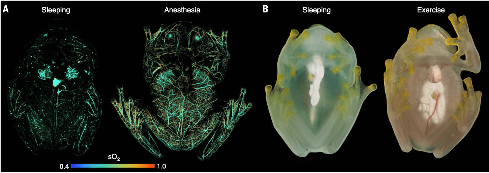
A glass frog hides its red blood cells while sleeping. (A) A high-resolution photoacoustic microscope image shows the red blood cells distribution within a frog while asleep and under anesthesia. (B) Flash photography shows the visible change in the frog while sleeping and awake.
The research team discovered that glass frogs can move nearly 90% of their visible red blood cells into their liver while resting or sleeping, which creates an optical effect close to invisibility, with clear plasma as the only fluid circulating in their bodies. Then, once they return to an alert state, the red blood cells move from the liver back into the rest of their body. For most living creatures, moving your entire blood supply into the liver would create an oxygen-starved state leading to dangerous clotting and organ damage, yet it produces no adverse effects for the frog.
The researchers noted that both frogs and humans share the same genetic and physiological factors that regulate hemostasis — the process of blood clot formation at the site of a vessel injury. As such, the results could prove invaluable for stroke and other vascular research.
Tracking the Vanishing Blood
In order to track these visible blood cells moving into the glass frog's liver, the research team harnessed the Zhou lab's piezoelectric high-frequency ultrasound transducer, fabricated by postdoctoral researcher Laiming Jiang. The transducer is a unique device that can capture and measure ultrasound waves for photoacoustic images.
"USC is a pioneer when it comes to this high-sensitivity high-frequency ultrasound device," Zhou said. "The principle of photoacoustic imaging is that we use a laser to shine into tissue. Then the tissue absorbs this laser light, and the blood temperature increases, producing a wave. Using our ultrasound transducer, we can receive this wave, which can generate the image."
Zhou said that his lab's device was able to produce high-frequency ultrasound over 30 up to 100 MHz, which can acquire a much higher resolution image than other forms of photoacoustic imaging.
The research team hopes that the study's findings may be harnessed to better understand the process of blood flow or to develop new anticoagulants or other cardiovascular drugs.
Published on February 21st, 2023
Last updated on February 21st, 2023


Comments
Post a Comment